
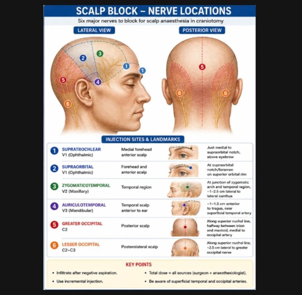
- Drugs used are
- Lignocaine 1.5% with adrenaline (Max 7 mg/kg)
- Bupivacaine 0.5% (Max dose 2mg/kg)
- Ropivacaine 0.75% (Max dose 3 mg/kg)
- Calculate the total local anaesthetic dose from all sources: Scalp block + pin infiltration + surgeon infiltration + subsequent top-ups
- The scalp is highly vascular and inadvertent intravascular injection can result in local anaesthetic systemic toxicity (LAST).
- Lidocaine IS useful for:Pin-site infiltration ,Additional local infiltration,Faster onset analgesia
- Ropivacaine is nowadays preferred over bupivacaine due to its lesser cardiotoxicity and its inherent vasoconstriction property.
ANAESTHETIC TECHNIQUES:
There is no recognized fixed anaesthetic regime followed for awake craniotomy.
Anaesthetic technique is often modified depending on the patient factors, pathology, tumour size, and its effect on the surrounding brain, length of surgery.
Most commonly followed ones (11) are:
- AWAKE-AWAKE-AWAKE
- ASLEEP-AWAKE-ASLEEP
- ASLEEP-AWAKE
- MAC
AWAKE-AWAKE-AWAKE (12,13)
Patients are awake throughout the procedure with or without minimal sedation.
Patients are maintained on spontaneous breathing without the use of an airway device.
The patients are awake during mapping and the surgical removal of pathological brain
ASLEEP-AWAKE-ASLEEP (4,13,14)
This technique involves induction of general anaesthesia and control of the airway with either a supraglottic device or an ETT.
When neurocognitive testing and intra-operative mapping needs to commence, the anaesthetic drugs are either reduced or stopped and the airway device is removed, when the patient has regained upper airway reflexes
Once resection of the lesion is complete, general anaesthesia can be re-introduced and with re-insertion of the airway device.
The airway device most often used are the Proseal LMA™, or intubating LMA, standard laryngeal mask (LMA) or Supreme LMA™, or even ETT is also used
ASLEEP-AWAKE:
This technique involves induction of general anaesthesia and control of the airway with either a supraglottic device or ETT.
When neurocognitive testing and intra operative mapping needs to commence, the anaesthetic drugs are either reduced or stopped and the airway device is removed.
Once resection of the lesion is complete, the sedation is restarted and patients are maintained on spontaneous respiration without using an airway device.
MAC: (4,5,15)
With MAC, the patient remains spontaneously breathing and does not routinely require an airway device.
The anaesthesiologist provides: Analgesia + Anxiolysis + Titrated sedation
while preserving: Airway reflexes + Spontaneous ventilation + Neurological cooperation.
SEDATION AGENTS: (4,5,15–17)
DRUG | BOLUS | INFUSION |
PROPOFOL | 0.5-1.0 mg/kg | Manual: 50-150 µg/kg/min TCI effect site (sedation): 2.4- 4.8mcg/ml TCI effect site (mapping): 0.6-1.2mcg/ml |
DEXMEDITOMEDINE | 0.5-1 µg/kg over 15 minutes | 0.2 -0.7 µg/kg/hr |
FENTANYL | 0.5-1µg/kg | 0.5 – 1 µg/ kg/hr |
REMIFENTANYL |
| Manual infusion: 0.03-0.05mcg/kg/min TCI effect site (sedation): 2-2.8ng/ml TCI effect site (mapping): 1.6-2ng/ml |
ALFENTANIL | 0.075mcg/kg | 0.0015mcg/kg/min |
SUFENTANIL | 7.5mcg/kg | 0.5mcg/kg/min |
MIDAZOLAM | 20-50 mcg/kg |
|
OTHER COMMONLY USED DRUGS:
THESE DRUGS ARE ADMINISTRED AT THE START OF THE CASE
- Inj. Paracetamol 15mg/kg – Analgesic
- Inj. Diclofenac 1.25 mg/kg – Analgesic
- Inj. Dexamethasone 8 mg -Anti edema effect
- Inj. Ondansetron 4 mg – For nausea and vomiting
- Inj. Glycopyrrolate 10mcg/kg – Antisialogue
- Inj. Levetiracetam 20mg/kg – Anticonvulsant
- Inj. Ceftriaxone 50mg/kg – Antibiotic
Challenges during Cortical Mapping and Surgical resection:
- Very important phase of the surgery
- All sedation is stopped well before testing
- Patient must be fully awake and cooperative especially when complex language and speech testing is planned.
- Patient may complain of pain during dura handling as it is very pain sensitive, so surgeons are asked to keep xylocaine-soaked gel pads over the dura before handling it to decrease the pain
- Intraoperative seizure can occur while doing the cortical mapping/stimulation, particularly in the vicinity of the motor cortex
- In case intraoperative seizures:
- Surgeon to be informed immediately and stimulation stopped
- Cold saline irrigation of the brain
- Maintain oxygenation and ventilation
- 1mg/kg Propofol bolus administered
- Small dose of thiopentone can also be tried
- If needed convert to GA if seizures not controlled
- Nausea and bradycardia may occur with deep cortical resection close to the midline, and this can be decreased by asking the surgeons to reduce the traction and administering anticholinergics.
- Functional mapping involves mainly the motor, language, speech area functions
- The exact testing battery should be determined jointly by the neurosurgical and neuropsychology teams according to the lesion and planned mapping strategy.(2,18,19)
Hand grip | Naming | Attention |
Finger movement | Reading | Memory |
Arm movement | Counting | Calculation |
Leg movement | Repetition | Executive Tasks |
Facial movement | Picture description |
|
| Spontaneous speech |
|

Intraoperative Complications:(1,4,20)
RESPIRATORY | CARDIOVASCULAR | NEUROLOGICAL | GENERAL |
Hypoventilation leading to hypoxia and hypercapnia | Hypotension during bolus administration of Propofol or dexmedetomidine | Seizures either focal or general | Nausea & Vomiting |
Malposition of SGA device | Hypertension during pinning or anxiety | Focal neurological deficit | Local Anaesthesia Systemic toxicity |
Aspiration | Bradycardia due to retraction or high dose of remifentanil | Tight brain /Brain swelling | Agitation/Restlessness |
Conversion to GA (Incidence -2%) | Hypertension due to increased adrenaline absorption | Venous air embolism especially if the head is above the heart | Delirium |
| Bradycardia/Tachycardia due to Trigeminocardiac reflex |
| Pain due to inadequate block or wearing off of the block |
Every awake craniotomy should have a pre-defined GA back up plan in case needed
Indications to convert to GA:
- Persistent airway compromise
- Severe respiratory depression
- Uncontrolled seizure
- Severe agitation
- Uncontrolled pain
- Severe nausea/vomiting
- Loss of cooperation
- Significant neurological deterioration
- Surgical requirement
POST OPERATIVE PERIOD:(4,6)
The success of awake craniotomies depends on patient cooperation along with the surgeon anaesthesiologist and IONM personnel/neuropsychologist.
Post-surgery assesses for
- Neurological status
- Consciousness
- Motor function
- Speech
- Pain
- Nausea/ Vomiting
- Airway and respiratory status
- Close neurological monitoring is required especially in the first 6hrs post-surgery as they may develop hematomas
- Always watch for any new onset deficits in immediate post-operative period
- Few patients may develop neurological deficits immediately after surgery despite nil intraoperative issues which is likely due to intraoperative handling and edema, such patients need reassurance that this is a transient deficit and will improve as the edema subsides
REFERENCES:
1. Kim SH, Choi SH. Anesthetic considerations for awake craniotomy. Anesthesiol Pain Med. 2020;15(3):269–74. doi:10.17085/apm.20050
2. Hervey-Jumper SL, Li J, Lau D, Molinaro AM, Perry DW, Meng L, et al. Awake craniotomy to maximize glioma resection: methods and technical nuances over a 27-year period. J Neurosurg. 2015;123(2):325–39. doi:10.3171/2014.10.JNS141520
3. Zhang JJY, Lee KS, Voisin MR, Hervey-Jumper SL. Awake craniotomy for resection of supratentorial glioblastoma: a systematic review and meta-analysis. Neuro-Oncol Adv. 2020;2(1):vdaa111. doi:10.1093/noajnl/vdaa111
4. Meng L, McDonagh DL, Berger MS, Gelb AW. Anesthesia for awake craniotomy: a how-to guide for the occasional practitioner. Can J Anesth. 2017;64(5):517–29. doi:10.1007/s12630-017-0840-1
5. Min KT. Practical guidance for monitored anesthesia care during awake craniotomy. Anesthesiol Pain Med. 2025;20(1):23–33. doi:10.17085/apm.24183
6. Guidelines Committee of the Japan Awake Surgery Conference. Guidelines for awake surgery. Neurol Med Chir (Tokyo). 2024;64(1):1–27. doi:10.2176/jns-nmc.2023-0111
7. Osborn I, Sebeo J. “Scalp block” during craniotomy: a classic technique revisited. J Neurosurg Anesthesiol. 2010;22(3):187–94.
8. Luo M, Zhao X, Deng M, Hu Y, Yang X, Mei Z, et al. Scalp nerve block, local anesthetic infiltration, and postoperative pain after craniotomy: a systematic review and network meta-analysis of randomized trials. J Neurosurg Anesthesiol. 2023;35(4):361–74. doi:10.1097/ANA.0000000000000868
9. Loukas M, Curry B, Jordan R, Wagner T, Tubbs RS, Cohen-Gadol AA. The innervation of the scalp: a comprehensive review including anatomy, pathology, and neurosurgical correlates. Surg Radiol Anat. 2008;30(8):675–82.
10. Kemp WJ, Tubbs RS, Cohen-Gadol AA. The innervation of the scalp: A comprehensive review including anatomy, pathology, and neurosurgical correlates. Surg Neurol Int. 2011 Dec 13;2:178. doi:10.4103/2152-7806.90699 PubMed PMID: 22276233; PubMed Central PMCID: PMC3262995.
11. Stevanovic A, Rossaint R, Veldeman M, Bilotta F, Coburn M. Anaesthesia management for awake craniotomy: systematic review and meta-analysis. PLoS ONE. 2016;11(5):e0156448. doi:10.1371/journal.pone.0156448
12. Hansen E, Seemann M, Zech N, Doenitz C, Luerding R, Brawanski A. Awake craniotomies without any sedation: the awake-awake-awake technique. Acta Neurochir (Wien). 2013;155(8):1417–24. doi:10.1007/s00701-013-1801-2
13. Seemann M, Zech N, Graf B, Hansen E. Anesthesiological management of awake craniotomy: asleep-awake-asleep technique or without sedation. Anaesthesist. 2015;64(2):128–36. doi:10.1007/s00101-014-2396-6
14. Sarang A, Dinsmore J. Anaesthesia for awake craniotomy–evolution of a technique that facilitates awake neurological testing. Br J Anaesth. 2003;90(2):161–5. doi:10.1093/bja/aeg037
15. Aghajanian S, Naeimi A, Mohammadifard F, Mohammadi I, Rajai Firouzabadi S, Baradaran Bagheri A, et al. Efficacy and safety of anesthetic agents in awake craniotomy using monitored anesthesia care protocol: a systematic review and meta-analysis. Neurosurg Rev. 2025;48(1):57. doi:10.1007/s10143-025-03176-y
16. Goettel N, Bharadwaj S, Venkatraghavan L, Mehta J, Bernstein M, Manninen PH. Dexmedetomidine vs propofol-remifentanil conscious sedation for awake craniotomy: a prospective randomized controlled trial. Br J Anaesth. 2016;116(6):811–21. doi:10.1093/bja/aew024
17. Akavipat P, Sookplung P, Lekprasert V, Kasemsiri C, Lerdsirisophon S. Dexmedetomidine for awake craniotomy: systematic review and meta-analysis. J Clin Neurosci. 2024;127:110765. doi:10.1016/j.jocn.2024.110765
18. Sattari SA, Rincon-Torroella J, Sattari AR, Feghali J, Yang W, Kim JE, et al. Awake versus asleep craniotomy for patients with eloquent glioma: a systematic review and meta-analysis. Neurosurgery. 2024;94(1):38–52. doi:10.1227/neu.0000000000002612
19. Collee E, Vincent A, Visch-Brink E, De Witte E, Dirven C, Satoer D. Localization patterns of speech and language errors during awake brain surgery: a systematic review. Neurosurg Rev. 2023;46:38. doi:10.1007/s10143-022-01943-9
20. Manninen PH, Tan TK. Postoperative nausea and vomiting after craniotomy for tumor surgery: a comparison between awake craniotomy and general anesthesia. J Clin Anesth. 2002;14(4):279–83. doi:10.1016/S0952-8180(02)00354-9