
ASTHMA EXACERBATION
Recommendations below are aligned primarily with the 2026 GINA Strategy Report, the current GINA reference available in 2026.
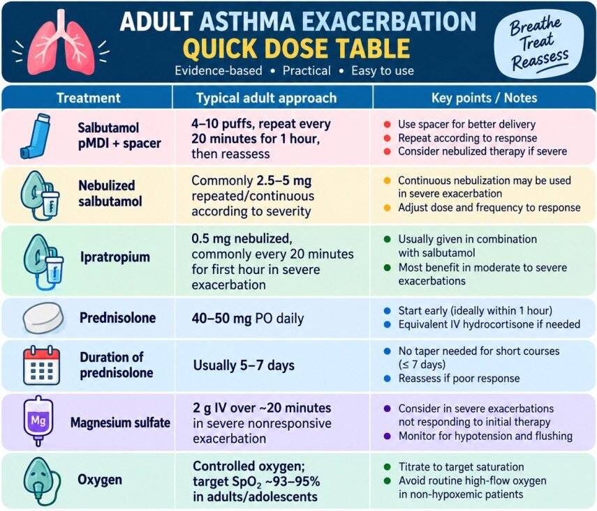
ADULT ASTHMA EXACERBATION — QUICK DOSE TABLE
Table of Contents
Toggle1. WHAT IS AN ASTHMA EXACERBATION?
An asthma exacerbation is an acute or subacute worsening of asthma symptoms and lung function compared with the patient’s usual state.Typical manifestations:
- Increasing dyspnea
- Wheezing
- Chest tightness
- Cough
- Increasing respiratory rate
- Increasing use of accessory muscles
- Falling PEF/FEV1
- Increasing requirement for reliever medication
- Hypoxemia in severe disease
Importantly, an exacerbation may occur in a patient with previously diagnosed asthma or may be the first presentation of asthma.
The terms “asthma attack,” “acute severe asthma,” “status asthmaticus,” and “severe exacerbation” are often used interchangeably clinically, but they are not perfectly synonymous.”Status asthmaticus” generally refers to: Severe asthma exacerbation that is persistent and refractory to conventional initial therapy.
2. PATHOPHYSIOLOGY
Trigger
↓
Airway inflammatory response
↓
Release of mediators:
- Histamine
- Leukotrienes
- Prostaglandins
- Cytokines
- Chemokines
↓
3 major airway abnormalities
1. Bronchial smooth-muscle contraction→ rapid airway narrowing
2. Airway mucosal edema→ further reduction in airway caliber
3. Excess mucus + mucus plugging→ partial/complete airway obstruction
↓
Increased airway resistance
↓
Expiratory flow limitation
↓
Air trapping
↓
Dynamic hyperinflation
↓
Increased work of breathing
↓
Respiratory muscle fatigue
↓
Ventilatory failure
Why EXPIRATORY FLOW IS MORE AFFECTED THAN INSPIRATORY FLOW
During expiration:
- Intrathoracic pressure rises
- Airways naturally become narrower
- Diseased small airways collapse more easily
- Expiratory flow becomes limited
The patient therefore has particular difficulty getting air out.
This creates:
Incomplete expiration → air trapping → End-expiratory lung volume progressively increases.This is:Dynamic hyperinflation or Auto-PEEP / intrinsic PEEP
WHY DYNAMIC HYPERINFLATION IS DANGEROUS
The diaphragm becomes flattened.Instead of being a dome:
Therefore respiratory muscles must generate much greater effort.
This creates:
More effort → more fatigue → worse ventilation → more CO₂ retention
It also produces important cardiovascular effects.
3. EFFECT OF HYPERINFLATION ON CARDIOVASCULAR SYSTEM
Severe hyperinflation:
Increases intrathoracic pressure
↓
Reduces venous return
↓
Reduces RV preload
↓
Reduces LV filling
↓
Potential reduction in cardiac output
Therefore a patient with severe asthma can become:
- Tachycardic
- Hypotensive
- Pulsus paradoxus positive
- Eventually peri-arrest
PULSUS PARADOXUS
Pulsus paradoxus = exaggerated inspiratory fall in systolic BP, classically:>10 mmHg
Mechanism
During inspiration:
↑ negative intrathoracic pressure—>↑ venous return to RV—>RV expands—>Interventricular septum shifts toward LV—>LV filling decreases—>LV stroke volume decreases—Inspiratory fall in systolic BP
4. WHY A NORMAL PaCO₂ CAN BE A WARNING SIGN
Early in an asthma attack:Respiratory rate increases
Therefore:PaCO₂ usually decreases.
But in a severely tachypneic asthmatic patient, a normal PaCO₂ can actually indicate worsening ventilatory failure.
Because that means
The patient is no longer able to increase alveolar ventilation due ton Increasing muscle fatigue
Therefore:In severe asthma:
Rising PaCO₂ = red flag
especially when associated with:
- Increasing fatigue
- Reduced air entry
- Altered mental status
- Increasing respiratory effort
- Falling pH
MIXED RESPIRATORY + METABOLIC ACIDOSIS
A very sick asthmatic patient may develop:
Respiratory acidosis + metabolic acidosis
Possible causes of metabolic acidosis:
- Lactic acidosis
- Hypoperfusion
- Respiratory muscle fatigue
- β₂-agonist-associated lactate elevation
- Sepsis
- Renal dysfunction
- Ketoacidosis
Thus:
pH may fall dramatically despite only moderate CO₂ elevation.
HOW SALBUTAMOL CAN INCREASE LACTATE
High-dose β₂-agonists can increase:
- Glycolysis
- Glycogenolysis
- Lipolysis
This increases pyruvate production and can increase lactate.
WHICH CAUSES tachypnea which may be mistaken for worsening bronchospasm.
5. COMMON PRECIPITANTS
Trigger Category | Examples / Clinical Significance |
A. Viral Respiratory Infections(Most common trigger overall) | Rhinovirus (most common), Influenza, RSV, SARS-CoV-2, and other respiratory viruses. Viral infections are the leading cause of acute asthma exacerbations in both children and adults. |
B. Allergen Exposure | House dust mites, pollens, animal dander, cockroach allergens, and mold spores. Exposure triggers IgE-mediated airway inflammation in sensitized individuals. |
C. Air Pollution | PM2.5, PM10, ozone, nitrogen dioxide, wildfire smoke, biomass smoke, and traffic-related pollution. Particularly important in urban environments and during periods of poor air quality. |
D. Tobacco Smoke | Active smoking, passive (secondhand) smoke exposure, and e-cigarette/vaping aerosols. Smoking reduces corticosteroid responsiveness and increases exacerbation frequency. |
E. Occupational Exposures | Flour dust, isocyanates, latex, industrial chemicals, wood dust, metal fumes, and cleaning agents. Consider occupational asthma if symptoms improve away from work. |
F. Drugs | β-blockers: Propranolol, timolol eye drops, metoprolol, atenolol (non-selective agents are highest risk). Aspirin/NSAIDs: Can precipitate attacks in susceptible patients, especially NSAID-exacerbated respiratory disease (N-ERD). |
G. Poor Adherence to ICS-Containing Therapy | One of the most important preventable causes of exacerbations. Reliance on bronchodilators without adequate inhaled corticosteroid therapy allows persistent airway inflammation. |
H. Incorrect Inhaler Technique | Medication fails to reach the lower airways despite apparent treatment adherence, making asthma appear “treatment resistant.” Inhaler technique should be checked at every visit. |
I. Psychological Stress | Stress and anxiety can worsen asthma control and trigger hyperventilation, but acute dyspnea should never be assumed to be anxiety until asthma and other serious causes are excluded. |
J. Exercise | Exercise-induced bronchoconstriction (EIB) typically occurs during or within 5–15 minutes after exercise, especially in cold, dry air. |
K. Gastroesophageal Reflux Disease (GERD) | GERD may worsen asthma symptoms through microaspiration and vagal reflexes in selected patients, particularly those with nocturnal asthma. |
6. CLINICAL PRESENTATION
Typical:
Symptoms
- Dyspnea
- Wheeze(A very severe asthmatic may actually have less wheeze because airflow is becoming extremely poor.)
- Chest tightness
- Cough
- Difficulty speaking
- Nocturnal symptoms
- Increased reliever requirement
Signs
- Tachypnea
- Tachycardia
- Wheezing
- Prolonged expiration
- Accessory muscle use
- Hyperinflation
- Reduced air entry
- Hypoxemia
SILENT CHEST
Silent chest = extremely dangerous.
It means:Airflow is so severely reduced that wheezing becomes minimal or absent.
GINA identifies drowsiness, confusion, or a silent chest as life-threatening features.
7. SEVERITY CLASSIFICATION
Feature | Mild–moderate | Severe | Life-threatening |
Speech | Phrases | Words | Unable to speak / very limited |
Position | May sit | Sitting hunched forward | Often unable to maintain position |
Mental status | Usually normal | Agitated | Drowsy/confused |
Respiratory rate | Increased | >30/min | Variable |
Accessory muscles | Usually absent | Present | Often marked/exhaustion |
Pulse | ~100–120/min | >120/min | May become bradycardic late |
SpO₂ on room air | 90–95% | <90% | Severe hypoxemia |
PEF | >50% predicted/best | ≤50% | Often unobtainable |
Chest | Wheeze | Loud wheeze | Silent chest |
GINA emphasizes using the worst clinical feature rather than relying on a single parameter.
DO NOT WAIT FOR PEF IN A VERY SICK PATIENT
PEF is useful when the patient can perform the maneuver.
But:
A severely distressed patient may not be able to generate a reliable PEF.
And ACEP notes that PEF monitoring has not been shown to improve outcomes or reliably predict admission when universally applied; its use should be individualized.
Therefore:Clinical assessment comes first.
Never delay lifesaving treatment just to obtain PEF.
8. Investigations
Investigation | When / Why to Perform |
Routine Laboratory Tests | Not required for every mild exacerbation. In moderate/severe or atypical disease, targeted investigations may be useful to identify complications, treatment-related abnormalities, or alternative diagnoses. |
Electrolytes | Particularly K⁺ and Mg²⁺ in moderate/severe attacks or after substantial β₂-agonist therapy. β₂-agonists can cause hypokalemia; hypomagnesemia may increase arrhythmia risk. |
Blood Glucose | Consider with systemic corticosteroid therapy, repeated/high-dose β₂-agonists, diabetes, or altered mental status. β₂-agonists and steroids can increase glucose levels. |
CBC | Not routine. Consider when bacterial infection, anemia, eosinophilia, or another alternative diagnosis is suspected. Leukocytosis alone does not prove bacterial infection, as β₂-agonists and corticosteroids can increase WBC count. |
Serum Lactate | Useful in patients receiving high-dose/repeated β₂-agonists, particularly when tachypnea persists or metabolic acidosis is unexplained. β₂-agonists can cause type B lactic acidosis, which may paradoxically increase respiratory drive despite improving bronchospasm. |
Chest X-ray (CXR) | Not routinely required in a typical asthma exacerbation. Consider when there is suspicion of pneumothorax, pneumonia, pneumomediastinum, foreign body, heart failure, focal chest findings, an alternative diagnosis, or unexpected clinical deterioration. |
ECG | Consider in older patients, severe/persistent tachycardia, chest pain, arrhythmia, significant β₂-agonist exposure, electrolyte abnormalities, or known cardiovascular disease. β₂-agonists may cause tachycardia, hypokalemia and, rarely, arrhythmias. |
ABG | Particularly useful in severe or life-threatening asthma. Consider with severe respiratory distress, altered mental status, suspected respiratory failure, silent chest, persistent hypoxemia, rising/normalizing PaCO₂ despite severe distress, or poor response to initial treatment. |
9. MANAGEMENT
OXYGEN
For adults/adolescents, GINA targets approximately:SpO₂ 93–95%
with adjustment for altitude where appropriate.If >50% FiO2 is required, strongly consider an alternative or additional diagnosis (e.g., pneumothorax, mucus plugging).
SALBUTAMOL
MDI + spacer
A common GINA regimen:4–10 puffs repeat:every 20 minutes for 1 hour then reassess.
Each standard salbutamol MDI puff is commonly:100 micrograms
Thus:
4 puffs = 400 micrograms
10 puffs = 1 mg nominal dose
Delivery with a spacer is important.
NEBULIZED SALBUTAMOL
Useful when:
- Patient cannot coordinate MDI
- Severe distress
- Unable to use spacer effectively
- Continuous/repeated nebulization is required
-Dose:2.5–5 mg nebulized Q20 minutes.
-In severe asthma continuous nebulized therapy(10-15 mg/hour initially)may be used under monitoring.
IV salbutamol
IV salbutamol may be considered as an exceptional rescue therapy in refractory, life-threatening asthma when adequate inhaled β₂-agonist delivery cannot be achieved or is ineffective, under ICU-level monitoring. It is not recommended routinely because evidence has not demonstrated benefit over intensive inhaled therapy and systemic toxicity is greater.
IPATROPIUM BROMIDE(SAMA)
- Add to SABA in:moderate-to-severe exacerbations, especially severe presentations or poor response to initial SABA.
- Typical adult nebulized dose:0.5 mg every 20 minutes for the first hour followed by 0.5 mg may be nebulized Q4 hours.
SYSTEMIC CORTICOSTEROIDS
Prednisolone 40–50 mg orally once daily usually for:
5–7 days for adults.
GINA recommends early systemic corticosteroids in moderate/severe exacerbations.
Steroids:
- Reduce airway inflammation
- Reduce mucosal edema
- Reduce inflammatory mediator activity
- Improve β₂-receptor responsiveness
- Reduce relapse
- Reduce hospitalization
IV VS ORAL STEROIDS
If the patient can swallow and absorb medication:
Oral corticosteroids are generally adequate.
IV therapy is reasonable when:
- Cannot swallow
- Severe vomiting
- Altered consciousness
- Need for parenteral therapy
Examples:
- Methylprednisolone IV
- Hydrocortisone 100 mg IV initially followed 50 mg IV every 6 hours
- DEXAMETHASONE 12–16 mg PO/IV once daily
Advantages:
- Long half-life
- Convenient dosing
- Potentially shorter course
But prednisolone remains a standard approach.
Hydrocortisone 20 mg ≈ prednisone 5 mg ≈ methylprednisolone 4 mg ≈ Dexamethasone 0.75 mg
MAGNESIUM SULFATE
Severe exacerbation with inadequate response to initial treatment.(use is controversial)
Dose:Magnesium sulfate 2 g IV given over 20 minutes.
Magnesium produces bronchodilation through several mechanisms, including:
- Calcium-channel effects
- Reduction in smooth-muscle contraction
- Modulation of acetylcholine release
Evidence for nebulized magnesium is less convincing than for selected IV use.
ADRENALINE / EPINEPHRINE
Not routine therapy for ordinary asthma exacerbation.
Use IM epinephrine when:
anaphylaxis or angioedema is present.
GINA explicitly recommends epinephrine first when the patient presents with features of anaphylaxis as well as asthma.
Typical adult anaphylaxis regimen:Epinephrine 0.5 mg IM
using 1 mg/mL (1:1000) solution
IV epinephrine may be considered only as an exceptional rescue therapy in refractory, life-threatening asthma when conventional inhaled therapy cannot be effectively delivered, and should be undertaken in a closely monitored critical-care setting. There is no standardized GINA-recommended IV epinephrine infusion dose for asthma exacerbation.
ANTIBIOTICS
Do NOT routinely give antibiotics.
Most exacerbations are not bacterial infections.
Consider antibiotics only when there is convincing evidence of bacterial infection, such as:
- Pneumonia
- Bacterial sinusitis in appropriate context
- Another documented bacterial infection
GINA specifically advises against routine antibiotic prescribing for asthma exacerbations.
THEOPHYLLINE
Not routinely recommended for acute asthma exacerbation.
Why?
Limited additional efficacy + significant toxicity.
Potential adverse effects:
- Nausea/vomiting
- Tremor
- Tachyarrhythmia
- Seizures
- Toxicity due to narrow therapeutic window
HELIOX
- Helium-oxygen mixtures reduce gas density.
- Formulated as a 70:30 or 80:20 mixture, so this isn’t an option for patients requiring >30% FiO2
- Theoretically:lower gas density → reduced turbulent flow resistance
- May help selected patients with severe airflow obstruction, but evidence is insufficient for routine use.
Montelukast
Montelukast 10 mg PO/NG once daily may be continued in an intubated adult who was already taking it chronically. It may also be considered as an adjunct in patients with aspirin/NSAID-exacerbated respiratory disease (AERD), although it is not established rescue therapy for the acute exacerbation.
β-BLOCKERS
Avoid non-selective β-blockers during acute severe bronchospasm when possible.
They can antagonize β₂-mediated bronchodilation.
If a cardiovascular indication makes β-blockade essential, specialist risk-benefit assessment is required.
10. SEDATION
Dexmedetomidine can provide sedation with relatively little respiratory depression, making it attractive in some spontaneously breathing/NIV situations.
However:
Major adverse effects
- Bradycardia
- Hypotension
Morphine should be avoided because histamine release could theoretically worsen bronchospasm
11. NONINVASIVE VENTILATION(NIV)
NIV/BiPAP may sometimes be considered in carefully selected severe asthma patients.
Potential theoretical benefits:
- Reduces work of breathing
- Provides inspiratory support
- May counter intrinsic PEEP
- Can improve ventilation
But evidence is considerably less robust than in COPD.
- The ideal respiratory rate might be around ~15-25 b/m.
- A respiratory rate >25 suggests excessive tachypnea, which may promote gas trapping.
- A respiratory rate <<15 raises the possibility of oversedation (especially following opioids).
HFNC in acute severe asthma
HFNC is not established as routine first-line respiratory support for acute asthma exacerbation. It can be considered in selected patients with hypoxemia or significant respiratory distress, but evidence in asthma is much weaker than in acute hypoxemic respiratory failure from other causes.patients who are unable to tolerate NIV or patients with less severe disease, HFNC can be used
At high HFNC flow:Nebulized aerosol + high gas flow
→ increased impaction/dilution
→ less aerosol reaching the distal airways.
12. INTUBATION
Intubation in severe asthma can precipitate:
- Hypotension
- Severe dynamic hyperinflation
- Barotrauma
- Cardiovascular collapse
- Arrest
INDICATIONS FOR INTUBATION
1. Cardiac/respiratory arrest
2. Severe altered mental status
3. Inability to protect airway
4. Severe exhaustion
5. Progressive respiratory acidosis
6. Refractory hypoxemia
7. Worsening despite maximal medical therapy
8. Severe hemodynamic instability
9. “Dying” clinical appearance
DO NOT INTUBATE BASED ON CO₂ ALONE
Procedure
KETAMINE
Ketamine is often attractive for induction in severe asthma because it:
- Provides sedation
- Provides analgesia
- Usually maintains cardiovascular tone relatively well
- Has bronchodilatory properties
Dose :1–2 mg/kg IV titrated to the clinical situation.
ETT
ETT size: In adults, use the largest practical ETT, commonly ≥8.0 mm internal diameter when anatomy permits. A larger tube reduces airway resistance and facilitates suctioning/bronchoscopy
⚠️ Avoid aggressive bag-mask ventilation
After induction/intubation, avoid delivering:
- High respiratory rates
- Excessive tidal volumes
- Excessively rapid manual ventilation
because:High minute ventilation→ inadequate expiratory time→ air trapping
13. VENTILATION STRATEGY
The biggest mistake is:Trying to normalize PaCO₂ rapidly.
This may require excessive minute ventilation.That causes:
short expiratory time
↓
air trapping
↓
dynamic hyperinflation
↓
hypotension/barotrauma
Therefore:Allow prolonged expiration.
Ventilator strategy | Rationale |
Low respiratory rate: ~8–12/min | Reduces minute ventilation and, most importantly, provides more time for expiration, minimizing dynamic hyperinflation/auto-PEEP. The rate should be individualized according to expiratory flow and degree of air trapping. |
Low-to-moderate tidal volume: ~6–8 mL/kg predicted body weight (PBW) | Provides lung-protective ventilation while avoiding excessive minute ventilation. Do not increase VT simply to normalize PaCO₂, as this can worsen hyperinflation. |
High inspiratory flow | Shortens inspiratory time and therefore increases available expiratory time. Commonly ~80–100 L/min, individualized according to airway pressures and flow curves. |
Long expiratory time | Aim for sufficient time for expiratory flow to return to zero before the next breath. An I:E ratio of approximately 1:3–1:5 or longer may be required in severe obstruction. The flow-time waveform is more useful than the ratio alone. |
Permissive hypercapnia | Accept an elevated PaCO₂ rather than increasing RR or VT aggressively to normalize it. This reduces dynamic hyperinflation and ventilator-induced injury. pH(maintain>7.2)and clinical status matter more than achieving a normal PaCO₂.Contraindications OF permissive hypercapnia:
|
Monitor for dynamic hyperinflation | Look for expiratory flow not returning to zero, rising plateau pressure, hypotension, worsening hyperinflation and increasing intrinsic PEEP (auto-PEEP). |
Neuromuscular blockade
- Avoid continuous paralytic infusions
- because patients often receive high-dose systemic corticosteroids, and prolonged corticosteroid exposure combined with neuromuscular blockade has been associated with ICU-acquired myopathy/weakness.
- Paralysis should therefore be reserved for selected situations, such as severe patient–ventilator dyssynchrony that persists despite adequate sedation(PROPOFOL/FENTANYL/KETAMINE), or life-threatening respiratory acidosis, hypoxemia or hemodynamic compromise where ventilator control cannot otherwise be achieved.
- If an NMBA is required, use the lowest effective dose for the shortest possible duration, provide adequate analgesia and deep sedation, and reassess frequently for discontinuation.
PEEP IN INTUBATED ASTHMA
FIRST MEASURE AUTO-PEEP(intrinsic PEEP)
During controlled ventilation:Use an:End-expiratory hold
The measured pressure can estimate:intrinsic PEEP, AutoPEEP makes difficult to trigger breath.
- If Patient is triggering the ventilator use 5 cm PEEP
- If Patient not Triggering Breath then External PEEP(PEEP that you set on ventilator) should be ~75% of the intrinsic PEEP.(Volume-cycled ventilation: Increasing PEEP causes an increase in plateau pressure.)
IMMEDIATE RESPONSE TO SEVERE AUTO-PEEP
An intubated asthmatic suddenly becomes:
- Hypotensive
- Tachycardic
- Difficult to ventilate
- High airway pressures
Think: Dynamic hyperinflation.
If the patient is crashing:Temporarily disconnect from ventilator
allow passive exhalation
while simultaneously assessing:
- Pneumothorax
- Tube obstruction
- Circuit problems
- Severe bronchospasm
If blood pressure rapidly improves after disconnecting, severe dynamic hyperinflation is strongly suspected.
This is a temporary emergency maneuver, not definitive treatment.
BAROTRAUMA
Severe asthma + mechanical ventilation can cause:
- Pneumothorax
- Pneumomediastinum
- Subcutaneous emphysema
Risk rises with:
- High airway pressures
- Severe air trapping
- Excessive minute ventilation
14. DIFFERENTIAL DIAGNOSIS
- COPD exacerbation
- Pulmonary edema
- Pulmonary embolism
- Pneumothorax
- Pneumonia
- Anaphylaxis
- Upper-airway obstruction(Wheeze loudest over throat.)
- Foreign body
- Vocal cord dysfunction / inducible laryngeal obstruction
- Aspirated material
- Metabolic acidosis with compensatory tachypnea
- Panic/hyperventilation
- Cardiac ischemia
15. ICU ADMISSION INDICATIONS
Consider ICU for:
- Life-threatening exacerbation
- Altered mental status
- Silent chest
- Respiratory failure
- Rising PaCO₂
- Severe acidosis
- Need for continuous nebulization
- NIV requiring close monitoring
- Mechanical ventilation
- Hemodynamic instability
- Failure of aggressive ED treatment
16. POSSIBLE DISCHARGE FEATURES
A patient is more suitable for discharge when:
- Symptoms have substantially improved
- Work of breathing is minimal
- Oxygenation is satisfactory on room air
- Reliever requirement has decreased
- PEF is improving
- PEF is generally >60–80% of personal best/predicted when feasible
- Patient can manage medications
- Home support is adequate
GINA uses these factors in its discharge assessment.
17. FOLLOW-UP
After an exacerbation, follow-up should be arranged relatively soon.
GINA recommends follow-up within approximately:
2–7 days for adults and earlier in children.
At follow-up assess:
- Symptoms
- Reliever use
- Lung function
- Adherence
- Inhaler technique
- Trigger exposure
- ICS therapy
- Action plan
- Need for specialist referral
18. WRITTEN ASTHMA ACTION PLAN
Every patient should ideally have an individualized written/digital action plan.
zone | What to do |
🟢 Green zone | Asthma is well controlled/stable. Minimal or no symptoms, normal activity, and reliever use is at usual/baseline level. Continue regular controller treatment as prescribed. |
🟡 Yellow zone | Asthma is worsening. Increasing cough, wheeze, breathlessness, chest tightness, night waking, reduced activity, or increased reliever requirement. Follow the written action plan for the prescribed temporary increase/step-up in treatment and reassess response. |
🔴 Red zone | Severe deterioration / severe exacerbation. Marked breathlessness, difficulty speaking, severe symptoms, rapidly worsening condition, poor response to reliever therapy, or features suggesting life-threatening asthma. Seek emergency medical care immediately and follow the emergency-treatment instructions in the action plan. |
Vaccination: Patients with asthma should receive routine age- and risk-appropriate vaccinations. Particular attention should be given to annual influenza vaccination, COVID-19 vaccination, and pneumococcal vaccination when indicated, because respiratory infections are important triggers of asthma exacerbations. Vaccination should generally be deferred only during moderate/severe acute illness;