
JVP(Jugular Venous Pressure)
INTRODUCTION
Topic | Key Point |
Jugular Venous Pulse (JVP) | • Oscillating top of vertical column of blood in right IJV that reflects pressure changes in Right Atrium in cardiac cycle. |
Jugular Venous Pressure (JVP) | • Vertical height of oscillating column of blood. |
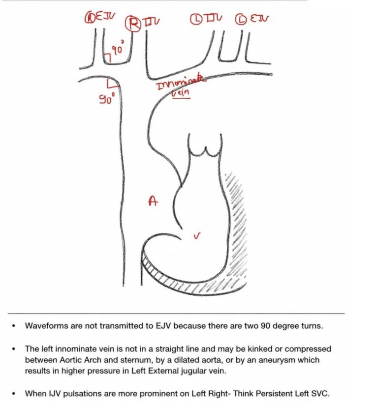
WHY RIGHT IJV OVER EJV ?
• IJV has a direct course to RA via Innominate Veins. |
• IJV is anatomically closer to RA. |
• IJV has no valves (EJV valves prevent transmission of RA pressure). |
• IJV reflects Right atrial pressure. |
• IJV shows Right atrial pressure and waveforms. |
CAROTID ARTERY VS JVP ?
Jugular venous pulsation | Carotid pulsation |
1. Superficial and lateral in the neck | Deeper and medial in the neck |
2. Better seen than felt | Better felt than seen |
3. Has two peaks and two troughs/cardiac cycle | Has single upstroke |
4. Descents > obvious than crests | Upstroke brisker and visible than descent |
5. x and y > prominent during inspiration | No effect |
6. a ↓ and v ↑ transiently during expiration | No effect |
7. Jugular venous pressure falls during inspiration | No effect |
8. Digital compression at the root of neck abolishes jugular venous pulse | No effect |

METHOD OF EXAMINATION
Position patient supine on an adjustable bed → Begin with 30° trunk elevation. |
Adjust trunk angle → Lower or elevate until maximum visible oscillations of the right internal jugular vein are seen. |
If JVP is low → Lower the trunk until pulsations become visible (reduces effect of gravity). |
If oscillations are not visible at 15° → Apply gentle abdominal compression with the flat of the hand → Increases venous return → Transiently reveals oscillations. |
If central venous pressure is high → Increase trunk elevation above the horizontal. |
Even then, the waves may be invincible. Careful inspection of the upper neck beneath the angle of jaw is important. |
If crests are not visible sitting bolt upright, gravity lowers venous column → Legs dangling pools blood in lower limb so venous return too heart over the side of the bed → If still not visible → Examine with the patient standing. |
EJV should also be analyzed in case of elevated venous pressure. |
Natural light is desirable for inspection. |
Flashlight or bed lamp can be used. |
Tangential lighting is best – Silhouette the neck veins to great advantage. |
The height of the venous column at the peak of the A wave and V waves generally is taken as an indication of the venous pressure. Although the actual mean jugular venous pressure will be slightly lower. |
ADDITIONAL CONSIDERATIONS, REFERENCE & MEASUREMENT
Head position → Neutral, slightly upward or downward and to the left → Avoid excessive upward tilt or leftward rotation, as these tense the right sternocleidomastoid and compress/obliterate the internal jugular pulse. |
Obese patients / short, thick neck → Adequate visualization of JVP is difficult. Inspiration / modest elevation of thorax brings out the venous waveforms. |
In tachycardia, A wave fuses with V wave → Carotid sinus pressure can be used to slow the heart rate. |
DTo analyze the waveform accurately, re-examine the JVP when the heart rate is slower. |
Reference point: Sternal angle of Louis. |
The crests of A and V waves (A prominent than V wave) do not exceed 3 cm above the angle of Louis when the trunk is 30° above horizontal. |
To measure RAP, add 5 cm H₂O (center of RA is 5 cm below sternal angle at 30–45° angle). |
1 mm Hg = 1.36 cm H₂O |
Timing of venous waves:1. Left carotid pulse carefully palpated with simultaneous visual inspection of right-sided jugular veins.2. Jugular venous pulse analyzed with auscultation of heart sounds. |
To analyze the waveform accurately, re-examine the JVP when the heart rate is slower. |
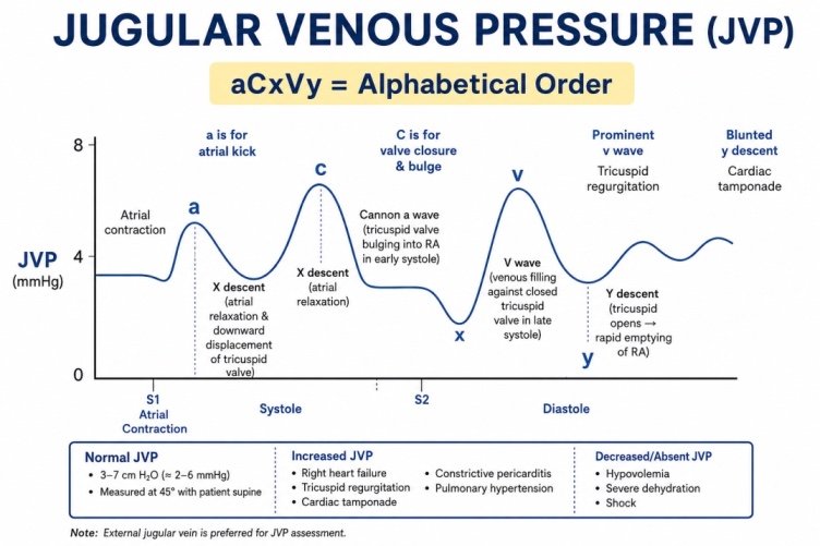
Table of Contents
ToggleJVP WAVEFORMS
Features | Causes |
a wave | Right atrial (RA) contraction |
x descent | Atrial relaxation |
x′ descent | RA floor descent and downward pulling of tricuspid valve (TV) by contracting right ventricle (RV) |
c wave | Impact of the carotid artery and upward bulging of the closed TV by contracting right ventricle (RV) |
v wave | RA filling during RV systole when TV is closed |
y descent | RA emptying during early RV diastole when TV opens |
Key Points
- As descents are more prominent than crests, prominence of descents helps distinguish the venous JVP from the arterial carotid pulse, which has a single brisk systolic upstroke.
- The x descent is usually the most prominent component of the normal JVP. The y descent is also prominent but is normally less deep than the x descent.
Sequence of JVP – A wave = S4 → S1 → X descent → S2 → V-wave peak → Y decent.
A WAVE
A wave | Mechanism – atrial contraction |
Elevated JVP – Most common | RV failure |
Causes of elevated JVP | 1. RV hypertrophy (↑ resistance to filling) / RVMI / RVCM2. Pulmonary stenosis3. Pulmonary hypertension / pulmonary embolism4. Tricuspid stenosis / tricuspid atresia / RA myxoma |
Bernheim effect | AS / HOCM |
Absent A wave | Atrial fibrillation |
Cannon A wave | Regular; JR / VT – 1:1 retrograde conduction |
Irregular cannon A wave | CHB / VPC / ventricular pacing |
V WAVE
Diminished V wave | Hypovolemia / Dehydration |
Prominent V wave – Causes |
|
X DESCENT – Descent of Floor of Right Atrium
X Descent – Prominent | Blunted X Descent |
• Cardiac tamponade | • Tricuspid regurgitation |
• Constrictive pericarditis | • Right atrial ischemia |
• RVMI |
|
• Restrictive cardiomyopathy |
|
• Atrial septal defect |
|
Y DESCENT
Prominent “y” descent | Absent “y” descent | Slow “y” descent |
• Constrictive pericarditis | • Cardiac tamponade | • Tricuspid stenosis |
• Severe tricuspid regurgitation | • Right ventricular infarction | • Right atrial myxoma |
• Severe RV failure | • Restrictive cardiomyopathy |
|
Constrictive pericarditis vs Cardiac Tamponade
Condition | X wave | Y wave |
Constrictive pericarditis | + | ++ (prominent Y) |
Cardiac tamponade | ++ (prominent X) | −− |
Tricuspid regurgitation (TR) | −− | ++ |
KUSSMAUL SIGN
Aspect | Details |
Definition | • Failure of decline in JVP during inspiration |
Associated Conditions | • Constrictive pericarditis/ Severe RHF / RVMI/ Restrictive cardiomyopathy/ Tricuspid stenosis |
TRICUSPID STENOSIS
Aspect | Details |
Prevalence | • Rare |
Classic Finding | • Classic cause of large A wave |
Clinical Pearl | • Think tricuspid stenosis in patients with large A wave in rheumatic mitral valve disease |
ABDOMINAL JUGULAR REFLUX
Principle | • Rise in jugular venous pressure (JVP) elicited by abdominal compression. |
Normal Site of Compression | • Right upper quadrant. |
Indications / Use | • In congestive hepatomegaly / central abdominal pressure, to evaluate JVP.• Preferred over hepatojugular reflux. |
Patient Preparation | • Patient relaxes and breathes quietly. |
Technique | • Examiner’s hand should be warm; contact gentle.• Firm compression is gradually increased until desired rise in JVP is observed.• Rapid pressure provokes reflex abdominal tension.• Minimize inadvertent straining (Valsalva manoeuvre).• Compress for 10–15 seconds (recent studies: 10 seconds adequate).• Historically: 15 seconds. |
Normal Response | • Transient rise of ~4 cm for about 4–5 cardiac cycles (~5 sec).• Initial increase followed by prompt fall. |
Positive (Sustained) Response | • Initial rise in JVP is not followed by prompt fall.• Rise is maintained throughout 10–15 seconds of compression. |
Clinical Significance of Positive Response | • Earliest sign of Right Heart Failure (RHF).• Also seen in: Tricuspid regurgitation (TR) / Constrictive pericarditis / Tamponade (TS) / Emphysema / Bronchospastic pulmonary disease. |
Negative (Absent) Response | • Obstruction / thrombosis of inferior vena cava (IVC) or hepatic veins (e.g., Budd–Chiari syndrome). |
Isolated LV Failure | • Response will be normal. |
Abdominal Jugular Reflex – False Positive Responses
- Severe COPD / pulmonary hyperinflation
- Increased sympathetic tone / Polycythemia
- Systemic venoconstriction
- Reduced venous-bed distensibility / Low venous capacitance
- Positive-pressure ventilation / High PEEP
- Markedly increased intra-abdominal pressure
- Pulmonary hypertension without clinical systemic congestion
For SuperSpecialists
JVP IN IWMI – INFERIOR WALL MYOCARDIAL INFARCTION
Situation | JVP Finding |
In IWMI: Hypotension + raised JVP + relatively clear lungs → think RV infarction | Elevated JVP |
RV infarction | Elevated JVP + prominent A wave + deep X & Y descents |
Prominent A wave | Impaired RV filling / reduced RV compliance |
Deep X & Y descents | May mimic constrictive pericarditis |
RV infarction with tricuspid valve damage → severe TR | Giant V waves |
Special Conditions | Key Features |
Superior vena cava obstruction | • Elevated JVP + no venous pulsation/waveforms are seen (elevated + non-pulsatile)+ Hepatojugular test negative + Venous collaterals visible in the upper chest |
Elevated JVP in person with normal cardiovascular system | • High cardiac output hyperkinetic state• Fluid overload• Obesity• Increased abdominal pressure• Asthma• Emphysema |
|
|
|
JVP in special situations | Details | |
TOF | • A wave may be normal or small. Tetralogy of Fallot: Systemic RV pressure → A wave remains normal because large VSD decompresses RV. Right atrial injury during surgery: ↓ A-wave amplitude postoperatively. | |
Atrial fibrillation (AF) | • A wave absent → RA fibrillation. X descent preserved, often reduced .• Annular descent is present, but atrial relaxation is absent .• V wave present → systemic veins empty into RA. | |
Ebstein anomaly | • JVP normal as RA size is very large. | |
CERVICAL VENOUS HUM
Nature | • Continuous benign murmur• Loudest in diastole |
Mechanism | • Mild compression of the internal jugular vein by the transverse process of atlas in patients with vigorous cardiac output |
Auscultation | • Right supraclavicular area / base of neck |
Position | • Only heard in sitting position• Disappears when subject is supine |
Maneuvers | • Valsalva maneuver / gentle compression of veins obliterates venous hum |
Differential | • May be confused with murmurs of PDA, AV fistula, aortic regurgitation• Can mimic carotid arterial bruit |
Clinical association | • Commonly occurs in patients undergoing hemodialysis |
JVP WITH SHOCK DIFFERENTIALS
Raised JVP + Shock |
• Congestive heart failure |
• Cardiac tamponade |
• Right ventricular infarction |
• Tension pneumothorax |
• Massive pulmonary embolism |
TR – CV COMPLEX
# | Finding | Detail |
1 | Absent x descent |
|
2 | Prominent systolic CV wave | Fusion of C + V waves, aka S wave |
3 | Rapid deep y descent | Due to rapid RA emptying |
4 | Correlates with | Severity of TR |
5 | Lancisi’s sign | Visible systolic earlobe pulsation with each heartbeat |
6 | AF + TR | Isolated giant V wave with rapid Y descent |
7 | Tall V wave | Called the systolic venous wave |
8 | Right-to-left head bob | Due to systolic expansion of right internal jugular vein |
9 | Mild TR | Venous contours normal at rest; abdominal compression / exaggerated inspiration / mild exercise brings out diagnostic V wave |
References
- Jonathan Abrams — Essentials of Cardiac Physical Diagnosis — Chapter 4: The Jugular Venous Pulse
- Braunwald’s Heart Disease — A Textbook of Cardiovascular Medicine — Chapter 13: History and Physical Examination: An Evidence-Based Approach — Jugular Venous Pressure and Waveform
- Perloff’s Physical Examination of the Heart and Circulation, 3rd ed. — Chapter 4: The Jugular and Peripheral Veins