
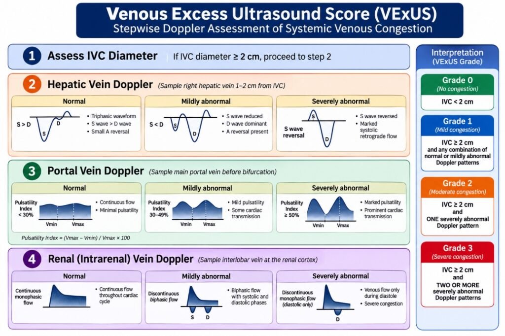
VExUS (Venous Excess Ultrasound Score)
VExUS is a point-of-care ultrasound (POCUS)-based scoring system used to quantify systemic venous congestionand predict organ dysfunction (especially congestive nephropathy) in critically ill patients.
It integrates:
- IVC size (macro congestion)
- Doppler patterns in venous systems (micro congestion transmission)
Table of Contents
ToggleWhy VExUS? (Core Concept)
Traditional markers (CVP, edema, weight gain) are:
- Late
- Non-specific
- Poor predictors of organ congestion
VExUS assesses backward failure (venous congestion):
Pathophysiological Cascade
↑ RA pressure → ↑ IVC pressure → transmission to:
- Hepatic veins → liver congestion
- Portal vein → splanchnic congestion
- Renal veins → ↓ GFR → AKI
This is congestive nephropathy
Although enthusiasm has grown in other aspects like sepsis, heart failure etc but systematic reviews conclude that evidence remains heterogeneous and further validation is needed before universal adoption.
VExUS Technique
- Position the patient supine (or slightly head-up if needed).
- Ask the patient to breathe normally.
- Locate the target vessel.
- Ask:“Breathe out normally… now hold your breath.”—measure at end-expiration(Reason- Primarily to reduce organ motion and obtain a stable Doppler signal,A short end-expiratory breath-hold minimizes respiratory effects so that the waveform primarily reflects the cardiac cycle, which is what VExUS evaluates.)
- Record 3–5 cardiac cycles (about 2–5 seconds).
- Resume normal breathing.
A. Inferior Vena Cava (IVC)
Measurement:
- Subcostal long-axis
- 1–2 cm from RA junction
Interpretation:
IVC | Meaning |
< 2 cm + collapsible | Low RA pressure |
> 2 cm + non-collapsible | High RA pressure |
VExUS requires dilated IVC (>2 cm) to proceed further(original VExUS protocol-Beaubien-Souligny)
But is this always true?No.
Clinical experience and later studies have identified situations where the IVC may be <2 cm but organ venous Doppler is still abnormal.
This is why many POCUS experts no longer rely exclusively on the IVC gatekeeper.
B. Hepatic Vein Doppler
Sample Right hepatic vein Near IVC—Pulse-wave Doppler
Normal Pattern(TRIPHASIC):S wave > D wave(forward flow)
followed by A wave
Congestion Pattern:
Pattern | Finding |
Mild | S < D |
Severe | S reversal |
Why hepatic vein changes first?
It drains directly into IVC.No capillary bed.Therefore First organ affected.
ANATOMY AND IDENTIFICATION
The portal triad contains:
- Portal vein
- Hepatic artery
- Common bile duct
Feature | Hepatic Vein | Portal Vein | Hepatic Artery |
Blood flow | Liver → IVC | Portal system → Liver | Aorta → Liver |
Direction | Away from liver | Towards liver | Towards liver |
Wall | Thin, indistinct | Thick echogenic wall | Thin, difficult to see |
Size | Largest | Large | Smallest |
PW Doppler | Triphasic | Continuous monophasic | Pulsatile arterial |
Respiratory variation | Marked | Mild | Minimal |
Cardiac pulsatility | Very marked | Minimal normally | Present |
Branching | Converge into IVC | Branch throughout liver | Travels with portal vein |
ROLE OF ECG
ECG event | Cardiac event | Hepatic vein Doppler |
P wave | Atrial contraction | A-wave reversal (small retrograde flow) |
QRS complex | Ventricular systole begins | S-wave starts(Systolic Forward Flow) |
T wave | End of systole | Transition to D-wave |
Early diastole | Tricuspid valve opens | D-wave(Diastolic Forward Flow) |
Without ECG, it can be difficult to determine whether the reversed wave is truly the systolic (S) wave or another component. ECG confirms its timing.
C. Portal Vein Doppler
Normal:Continuous, monophasic flow
Abnormal:Pulsatile flow (due to RA pressure transmission)
Portal Pulsatility Fraction (PPF):
PPF=(Vmax −Vmin)/Vmax ×100
PPF | Severity |
< 30% | Normal |
30–50% | Mild |
> 50% | Severe |
D. Intrarenal Vein Doppler
Obtained from Interlobar vein
Normal:Continuous venous flow
Congestion:
Pattern | Interpretation |
Biphasic | Moderate |
Monophasic (discontinuous) | Severe |
Direct reflection of renal venous hypertension
VExUS Grading System
Only applied when IVC is dilated
Grade | Criteria |
Grade 0 | IVC normal |
Grade 1 | IVC dilated + mild Doppler changes |
Grade 2 | IVC dilated + 1 severe abnormal Doppler |
Grade 3 | IVC dilated + ≥2 severe Doppler abnormalities |
Severe abnormalities:
- Hepatic vein → S reversal
- Portal vein → PPF > 50%
- Renal vein → monophasic flow
Clinical Significance
A. Acute Kidney Injury (AKI)
- Strong predictor of congestive AKI
- Better than CVP
Especially in:
- Post-cardiac surgery
- Septic patients with fluid overload
B. Fluid Management in ICU
Scenario | Role of VExUS |
Fluid resuscitation | Avoid over-resuscitation |
Shock | Differentiate hypovolemia vs congestion |
De-resuscitation | Guide diuretics/ultrafiltration |
Grade | Management |
0–1 | Fluids if needed |
2 | Caution → avoid overload |
3 | Active decongestion (diuretics/CRRT) |
C. Heart Failure
- Detect subclinical congestion
- Guide diuretic therapy
D. Perioperative Medicine
- Predict post-op AKI
- Optimize fluid balance
Limitations
- Not a direct measure of volume status.
- Elevated RAP from severe TR, pulmonary hypertension, RV dysfunction, or mechanical ventilation can alter waveforms independent of excess body fluid.
- Portal hypertension and chronic liver disease may confound portal vein interpretation.
- Technically challenging in obesity, abdominal wounds, dressings, bowel gas, or poor acoustic windows.
- Requires training in Doppler acquisition and interpretation.
- Should never be used in isolation to decide on fluids or diuretics; integrate with clinical examination, echocardiography, lung ultrasound, perfusion markers, urine output, and laboratory data.