
Inter scalene block
Brief history:
First brachial plexus block was performed by William Steward Halsted in 1889 → by exposing the brachial plexus and using cocaine for blockade.
Inter-scalene block was first described by Winnie in 1970.
It often spares the lower branches of the plexus the C8 and T1 fibres innervating ulnar border of the forearm.
Table of Contents
ToggleCLINICAL IMPLICATION –
Low inter-scalene block below C6 just superior to clavicle was devised to provide sufficient anaesthesia and analgesia for procedures on the lower part of upper arm.
Relevant anatomy:
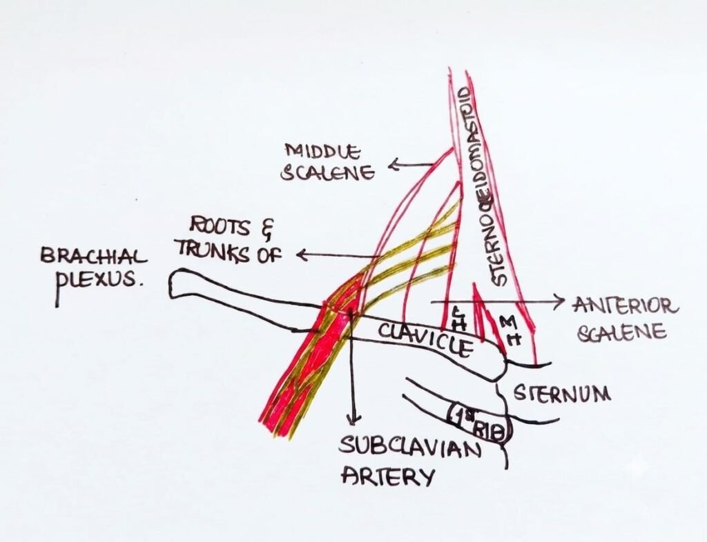
After leaving the inter-vertebral foramina, the nerves course antero- laterally and inferiorly to lie between the anterior and middle scalene muscles and fuse literally to enclose the brachial plexus in a fascial sheath.
Anatomical factors helpful in successful blockade:
- The fascial peri-vascular sheath – extends from the origin the scalene muscles to the middle of upper arm.
- The vertical arrangement of cervical nerve roots which form the trunks of brachial plexus
Indications:
- Anaesthesia and analgesia for open and shoulder surgery.(eg: acromioplasties, total shoulder replacement, debridement of labral and rotator cuff tears)
- Mobilisation (eg: frozen shoulder)
- Closed reduction of the shoulder dislocation
- Anaesthesia for upper arm surgery
PATIENT POSITIONING AND ANATOMICAL LANDMARKS
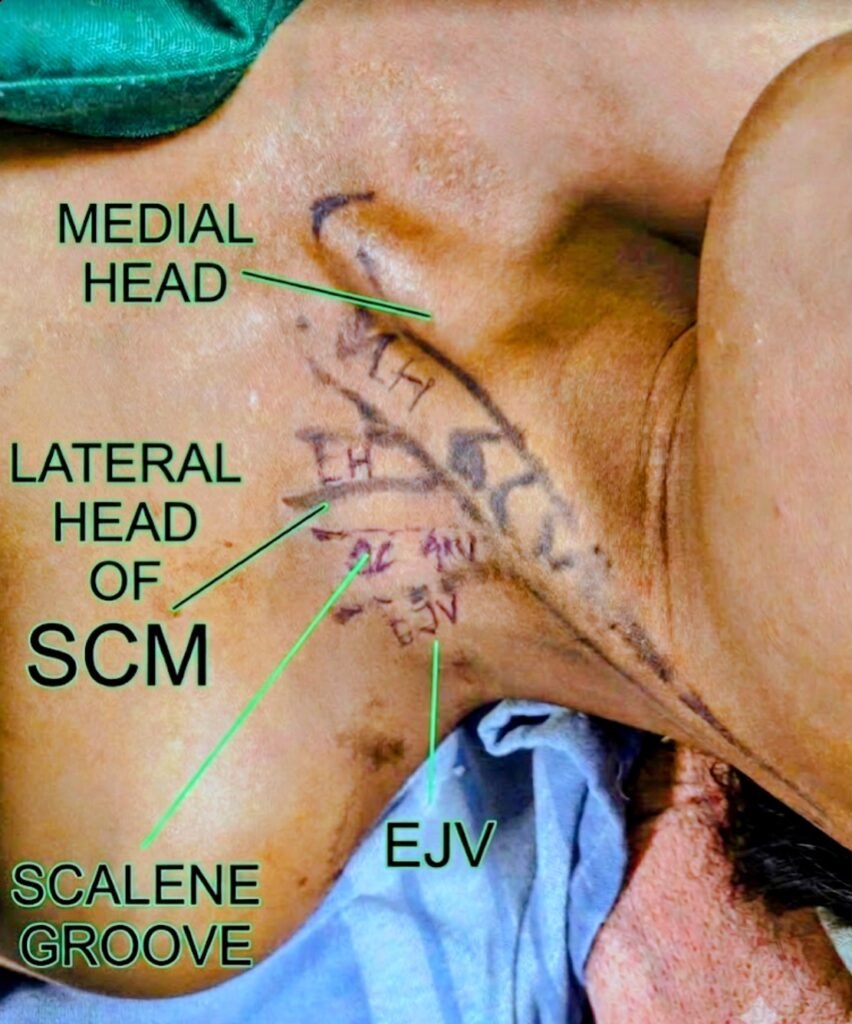
Patient is made to lie down and raise his head against closed glottis
This helps to make sternocleidomastoid muscle prominent.
3 anatomical landmarks are most significant
- Posterior border of sternocleidomastoid
- External jugular vein
- Scalene groove
- Equipments
- Standard regional anaesthesia tray
- Sterile towels for draping
- Sterile gauze pieces
- 2 of 10/20 mL syringes containing local anaesthetic
- Sterile gloves
- Marking pen
- Surface electrodes
- 3 mL syringe and 25 G needle for skin infiltration with local anaesthetic
- Peripheral nerve stimulator with stimuplex needle
- usg machine
Techniques of inter-scalene block
- Landmark guided
- PNS guided
- USG guided
Landmark guided Inter-scalene block
- Skin overlying the area is prepared by antiseptic solution
- At the level of cricothyroid notch (C6 ) at the inter-scalene groove, a 25 gauge,11/2 inch needle is inserted with a slight caudad and inferior trajectory
- If inter-scalene groove is not identified , the needle is inserted slightly behind posterior border of sternocleidomastoid.
PARASTHESIA will occur at 1/4 to 1 inch depth when needle is advanced quite slowly in perpendicular direction to reach inter-scalene groove and it’s tip impinges the brachial plexus.
Gentle aspiration and if aspiration is negative , local anaesthetic is injected slowly with close monitoring of the patient.
Nerve stimulation technique
Same as landmark technique using 22G 35 -50 mm insulated needle directed medial, caudal and posterior in the direction of C6 transverse process near posterior border of sternocleidomastoid corresponding to cricothyroid.
Caudal tilt is important as it avoids-
- Either entering neural foramen
- Injection into the dural nerve root sheath
Thus preventing spinal cord injury and high spinal .
Nerve localisation–
Apply an initial current of 0.8mA – 1mA and the current is aimed for a threshold of 0.4 mA before injecting after motor response.
Avoid diaphragm /trapezius stitches with cervical plexus stimulation.
Response should be from→pectoralis muscle, deltoid muscle, triceps muscle, biceps muscle or any twitch of the muscle of the hand or the fore-arm.
After negative aspiration gradually 20 to 30 mL of local anaesthetic is injected in small increments
(Picture 3)
USG guided technique
Advantages
- Distribution of local anaesthetic is visualised for adequate spread around the plexus
- Allows multiple injections around the brachial plexus thus eliminating the possibility of failed block
- Helps to reduce the volume of anaesthetics
- Avoids the risk of major vessel or nerve puncture
- Repetition of block is also possible
A linear USG probe is placed over previously identified location ( same as the one used in landmark guided and PNS techniques) in the transverse plane and a sonogram is obtained.
At inter-scalene level structures observed are:
- Superficially — pre-vertebral fascia, superficial cervical plexus and sternocleidomastoid muscle are observed
- Medially — carotid artery, anterior scalene muscle
- Laterally — middle scalene muscle
- Transducer is moved in superior – inferior direction until 2/more brachial plexus trunks are seen in the space between scalenes.
- In plane approach preferred.
Sonoanatomy
Roots of brachial plexus appear as hypoechoeic round or oval multifascicular structures with hyperechoeic perineurium
Colour Doppler used to identify carotid artery,internal jugular vein and vertebral artery
Probe is moved slightly superior and inferiorly so as to get the appearance of traffic spotlight sign of brachial plexus
Probe settings
Depth 3cm, frequency 8-14 Hz, linear transducer
Patient position
Supine with head turned away from side to be blocked
Transducer position
Transverse orientation on the neck over the pulse of carotid artery 4 cm above clavicle
Slide the transducer laterally and slightly up and down to visualise traffic signal sign .
In plane approach used to inject local anaesthetic
Tips –
- Higher resistance to injection >15psi pressure indicates intra fascicular injection and requiring repositioning of needle.
- Gentle pressure with transducer prevents collapse of vein and their subsequent puncture
Complications
- Vascular puncture
- Hematoma
- Infection
- Local anaesthetic systemic toxicity
- Nerve injury
- Total spinal anaesthesia
- Horne syndrome
- Diaphragm paralysis
- Epidural injection
- Pneumothorax
Contra – indications
- Contralateral phrenic and recurrent laryngeal nerve paresis
- Chronic obstructive pulmonary disease (relative contraindication)
General tips
- General neurological examination to identify pre-existing neurological deficits is necessary before brachial plexus block so that such effects are not later attributed to the block
- Supplementation for C8 fibers may be needed by peripherally blocking ulnar nerve in surgeries where C8 distribution is involved which may get spared with inter scalene block
References
Millers anaesthesia 8th edition
Textbook of anaesthesia Barash 7th edition
Hadzics regional anaesthesia 5th edition
Waldmann’s interventional pain management 5th edition
Nysora website