
Supraclavicular brachial plexus block
History
First introduced by Kulemkempff and Percy in 1911 for mid humerus level to hand surgeries.
Relevant anatomy
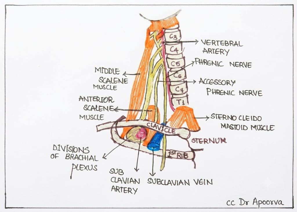
- Targets the trunks of brachial plexus formed by C5 -T1 when they form divisions at the level of clavicle.
- Subclavian artery lies on top of fist rib along with brachial plexus lying postero-medial to the subclavian artery.
- Subclavian vein and the scalenus anterior muscle are medial to the subclavian artery.
- Apex of pleura extends into the base of the neck above medial 1/3 rd of the clavicle.
- Phrenic nerve passes behind subclavian vein.
Clinical implications
- Brachial plexus confined to small area within peri vascular sheath here with densely packed divisions leading to dense blockade of the entire sensory, motor and sympathetic innervation of upper extremity
- Excluding uppermost part of medial aspect of forearm except T2 distribution of uppermost part of medial aspect of the arm.
- One has to be careful about this structure as phrenic palsy may occur with supraclavicular block if needle is inserted medially.
Indications
- Anaesthesia and analgesia of the entire upper extremity distal to shoulder including upper arm,elbow,forearm, wrist and hand.
- Diagnostic tool for upper limb pain evaluation.
- Treatment of chronic pain conditions like herpes zoster ,acute brachial plexus neuritis, trauma to upper limb,reflex sympathetic dystrophy of upper limb, cancer pain.

Surface marking and patient positioning
- Patient is made to lie down supine or semi recumbent with head turned to opposite side to be blocked.
- Sand bag could be used behind shoulder for better access the space.
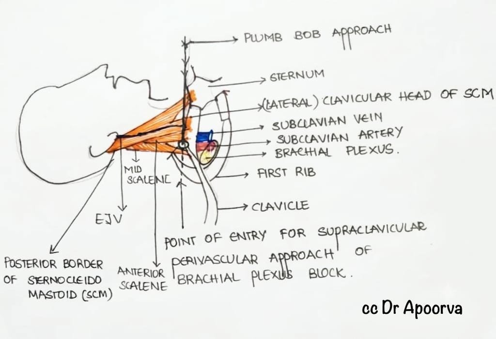
3 important landmarks are
- Posterior border of sternocleidomastoid muscle
- Mid point of clavicle
- Mark an X on inter-scalene groove just above the clavicle and behind about 1cm
Pulsation of subclavian artery above clavicle lateral to posterior border of sternocleidomastoid
Techniques
- Paresthesia method using landmarks
- PNS guided
- USG Guided
Landmark guided technique
- The perpendicular trajectory shown in picture depicts the masons plumb bob approach .
- Needle advanced very slowly until paresthesia is elicited at about 3/4th or 1 inch .
- If first rib is hit before eliciting parasthesia walk over the rib laterally until paresthesia is elicited
- Tip: medial advancement of needle leads to pneumothorax.
PNS guided technique
- Positioning same as landmark approach
- Entry point of stimulator needle same as described before for landmark guided approach about 2.5 cm lateral to lateral head of sternocleidomastoïd plexus can be present.
- Palpate for subclavian artery at this spot, needle insertion will be immediate cephalad to palpating finger.
- Nerve stimulator is connected to stimuplex needle and set to deliver 0.8-1mA current at 1 Hz frequency and 0.1ms of pulse duration.
- Needle entry-Mason’s plump bob approach with slight caudal orientation → advance needles→ upper trunk muscle twitch elicited at shoulder muscles-→ if no upper trunk twitch after advancing needle 1 cm of depth advance needle caudal to palpating finger with slight posterior angle.
- Needle at middle trunk biceps, triceps, pectoralis twitch→ advance towards lower trunk leads to finger twitch.
- → Injection after gentle aspiration → when motor response to fingers present at 0.5 mA → avoid injection at high resistance >15 psi
USG guided technique
- Patient is made to lie supine with head to opposite side to be blocked
- Operator stands or sits on side of the block
- USG machine is placed on opposite side to be blocked so that patient operator and machine as in single straight line
- 45 degree head up can be used for facilitating the block
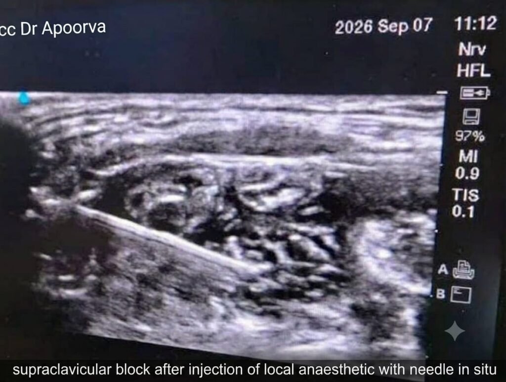
- High frequency linear probe is used to visualise the structures and the transducer is places transversely over the supraclavicular fossa in coronal oblique plane and image is optimised by tilting ,rotating and sliding the transducer probe.
Sonoanatomy
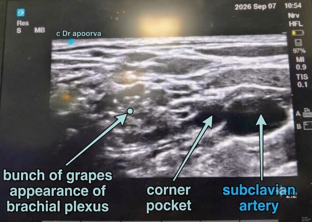
- Subclavian artery – round hypo echoic non compressible and pulsatile →medially
- First rib -bright hyper echoic with acoustic shadow → inferiorly
- Brachial plexus – BUNCH OF GRAPES multiple hypo echoic structures with hyper echoic rim → superolaterally
- These 3 constitute corner pocket.
- Pleura- hyper echoic similar to first rib but with lung sliding sign
- Scalene muscle – hyper echoic at insertion to first rib
- Thyro cervical trunk and transverse cervical artery – small pulsatile structures obscured by subclavian vessel cam be identified by colour Doppler
- Needle is inserted in plane lateral to medial to probe at the area of bunch of grapes with subclavian artery medial to plexus and on top of first rib
- Subclavian vein being medial to scalene anterior
- First the 8 ball or corner pocket which is USG landmark where C8 fibers of ulnar nerve is located is targeted later rest of the plexus covered
- Dual guidance technique with nerve stimulation enables successful block effectively.
Tips
- For tourniquet supplement with intercostobrachial block
- Injection of local anaesthetic medial and anterior to subclavian artery causes phrenic nerve palsy
- If corner pocket is missed supplement with ulnar nerve block distally
- Fanning the probe improves USG visibility
- To improve intervention space use sandbag below shoulder on side of block
- Use colour Doppler to rule out presence of transverse cervical artery.
Complications
- Hematoma
- Pneumothorax
- Epidural injection
- Vertebral artery injection
- Variable ipsilateral phrenic nerve block
- Subarachnoid injection
- Horners syndrome
- Spread of anaesthetics to other tissues causing ipsilateral numbness of face,recurrent laryngeal nerve palsy leading to variable paralysis of vocal cord.
- Local anaesthetic systemic toxicity
Contraindications
- Local infection
- On anti coagulation
- Severe systemic coagulopathy
References
- Millers anaesthesia 8th edition
- Barash textbook of anaesthesia 7th edition
- Hadzics Regional anaesthesia and pain management 2nd edition
- Manoj K Karmakar Atlas of sonoanatomy for regional anaesthesia and pain management 1st edition
- Waldmanns interventional pain management 5th edition
- Nysora website