
AIRWAY ASSESMENT
Objectives of airway assessment
- Identify a potentially difficult airway.
- Anticipate the type of difficulty likely to occur.
- Prepare appropriate equipment and personnel.
- Develop a primary and alternative airway plan.
- Maintain oxygenation throughout airway management.
- Reduce airway trauma and complications.
- Anticipate difficulty with extubation and reintubation.
No single airway examination test can reliably predict all difficult airways.
Table of Contents
ToggleHISTORY
Previous Anaesthetic History
One of the most important components of airway assessment is asking about previous anaesthesia.
Specifically ask about:
- Previous difficult intubation
- Difficult mask ventilation
- Difficult supraglottic airway placement
- Multiple intubation attempts
- Failed intubation
- Previous awake intubation
- Airway trauma
- Dental injury
- Aspiration
- Difficult extubation
Medical History
Craniofacial abnormalities | Upper airway pathology | Conditions affecting cervical spine movement | Other important conditions |
Micrognathia | Supraglottic tumours | Cervical spine instability | Acromegaly |
Retrognathia | Laryngeal tumours | Rheumatoid arthritis | Obstructive sleep apnoea |
Pierre Robin sequence | Airway oedema | Ankylosing spondylitis | Morbid obesity |
Treacher Collins syndrome | Epiglottitis | Cervical spine trauma | Previous head and neck surgery |
Goldenhar syndrome | Peritonsillar abscess | Previous cervical spine sur | Neck radiation |
Down syndrome | Retropharyngeal abscess |
| Burns and contractures |
Other congenital craniofacial abnormalities | Ludwig’s angina |
| Thyroid/neck masses |
| Foreign body |
|
|
| Airway stenosis |
|
|
LOCAL AIRWAY EXAMINATION
FACE | NOSE | TEETH | ORAL CAVITY | NECK |
Micrognathia | Nasal deformity | Loose teeth | Mouth opening | Neck circumference |
Retrognathia | Nasal patency | Prominent/buck teeth | Tongue size | Neck length |
Facial trauma |
| Protruding incisors | Palate | Neck mobility |
Craniofacial abnormalities |
| Crowns and bridges | Mandibular size | Masses |
Burns/contractures |
| Dentures | Oropharyngeal space | Scars |
|
| Poor dentition | Masses or lesions | Radiation changes |
|
|
| Mandibular protrusion | Cervical spine abnormalities |
Airway Measurements
Measurement | How measured | Significance |
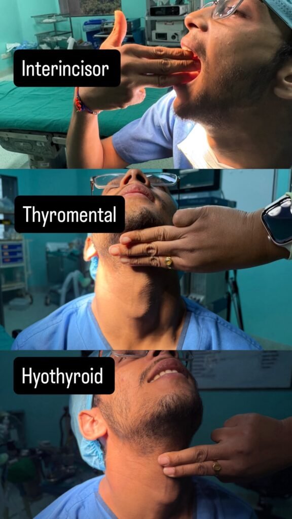
Mouth opening | Inter-incisor distance | Reduced opening may restrict insertion and manipulation of airway devices |
Thyromental distance | Mentum → thyroid notch with neck extended | Short distance suggests reduced mandibular space |
Hyomental distance | Hyoid bone → mentum, usually with the neck in a neutral position | Short distance may indicate reduced submandibular space and increased difficulty with airway management |
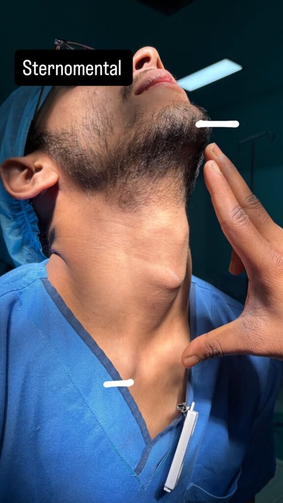
Sternomental distance | Mentum → sternal notch with neck extended | Gives information about neck length and extension |
Neck mobility | Flexion/extension | Reduced movement may make laryngoscopy difficult |
Mandibular protrusion | Ability of lower incisors to move beyond upper incisors | Assesses mandibular mobility |
Approximate values used in airway assessment
Parameter | Favourable | Concerning |
Mouth opening | >4 cm | <3 cm |
Thyromental distance | >6.5 cm | <6 cm |
Hyomental distance | >4 cm | <4 cm |
Sternomental distance | >12.5 cm | <12 cm |
Note: These are approximate clinical thresholds and should not be used in isolation.
Airway assessment TESTS
Always better to use a combination of tests
- Oropharynx
- Atlanto-Occipital Joint
- Submandibular Space
- Radiological
Mallampati Classification
Class | Structures visible | Interpretation |
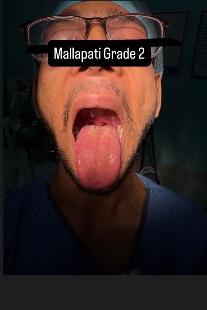
I | Soft palate, fauces, uvula and pillars | Generally favourable |
II | Soft palate, fauces and part/base of uvula | Generally favourable |
III | Soft palate and base of uvula | Increased likelihood of difficult laryngoscopy |
IV | Hard palate only | Increased likelihood of difficult laryngoscopy |
Important point
Mallampati is a predictor, not a diagnosis of difficult intubation.
It should always be interpreted together with other airway findings
Cormack–Lehane Classification
NOT a preoperative examination; it is an intraoperative laryngoscopic view
Grade | Laryngoscopic view |
Grade I | Full view of glottis |
Grade II | Partial view of glottis |
Grade III | Epiglottis visible but glottis not visible |
Grade IV | Neither glottis nor epiglottis visible |
Modified classification
Grade | Description |
IIa | Partial view of glottis |
IIb | Only posterior glottic structures/arytenoids visible |
IIIa | Epiglottis visible and can be lifted |
IIIb | Epiglottis visible but cannot be lifted |
Important distinction
Test | What it tells you |
Mallampati | Preoperative airway assessment |
Cormack–Lehane | Laryngoscopic view obtained during direct laryngoscopy |
Temporomandibular Joint (TMJ) & Mandibular Examination
The TMJ and mandible are assessed because adequate mouth opening and mandibular mobility are important for insertion and manipulation of the laryngoscope and for achieving a good laryngeal view.
Examination / Test | Technique | Normal / Expected Finding | Significance |
Inter-incisor distance (mouth opening) | Ask the patient to open the mouth maximally. Measure the distance between the upper and lower incisors. | Usually ≥ 3.5–4 cm | Reduced mouth opening may make insertion and manipulation of the laryngoscope difficult. |
Mandibular protrusion | Ask the patient to move the lower jaw as far forward as possible. | Good forward movement of the mandible | Assesses the range of anterior mandibular movement. Restricted protrusion may be associated with difficult laryngoscopy. |
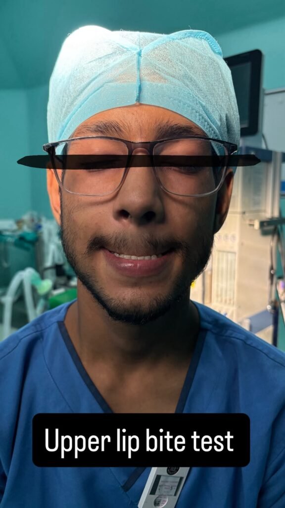
Upper Lip Bite Test (ULBT) | Ask the patient to bite the upper lip with the lower incisors. | Class I: lower incisors can bite the upper lip above the vermilion line | Assesses mandibular mobility and dental relationship. Higher classes are associated with increased difficulty. |
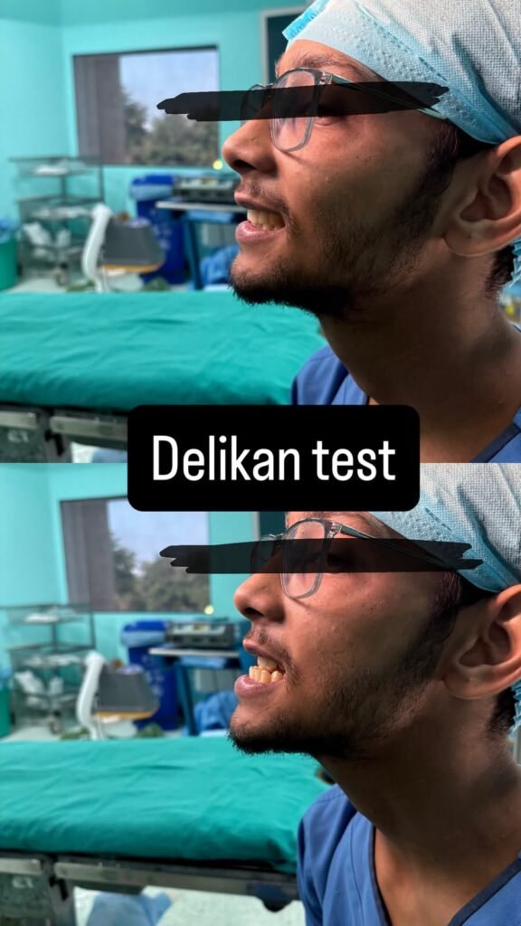
Delikan test | Ask the patient to protrude the mandible and assess whether the lower incisors can move in front of the upper incisors. | Lower incisors can protrude beyond the upper incisors | Specifically assesses the ability of the mandible to advance anteriorly relative to the maxilla. |
TMJ movement | Assess mandibular opening, protrusion and lateral movements. Observe for pain, clicking or restriction. | Full, symmetrical movement without significant restriction | TMJ restriction can reduce mouth opening and mandibular mobility. |
Mandibular morphology | Inspect for micrognathia, retrognathia, receding mandible and prominent incisors. | Adequate mandibular size and projection | Small or receding mandible reduces the available submandibular space and may make laryngoscopy difficult. |
Are mandibular protrusion and the Delikan test the same?
They are closely related but are not exactly the same.
- Mandibular protrusion is a general assessment of how far the patient can move the lower jaw forward.
- The Delikan test is a specific assessment of the relationship between the lower and upper incisors during mandibular protrusion—specifically, whether the lower incisors can move in front of the upper incisors.
Therefore, mandibular protrusion describes the movement, whereas the Delikan test assesses the resulting anterior dental relationship.
Key distinction
- ULBT: assesses whether the lower incisors can bite the upper lip.
- Delikan test: assesses whether the lower incisors can protrude beyond the upper incisors.
- Mandibular protrusion: assesses the overall forward movement of the mandible.
Upper Lip Bite Test
Class | Finding |
Class I | Lower incisors can bite above the vermilion border of the upper lip |
Class II | Lower incisors can bite the upper lip but not above the vermilion border |
Class III | Lower incisors cannot reach the upper lip |
Significance: Assesses mandibular mobility and ability to subluxate the mandible.Submandibular Space Examination
The submandibular space is extremely important because the tongue must be displaced into this space during direct laryngoscopy.
A small or poorly compliant submandibular space can make tongue displacement difficult.
Examination / Test | How assessed | Significance |
Thyromental distance (TMD) | Measure from mentum to thyroid notch with neck extended | Estimates available submandibular space |
Sternomental distance (SMD) | Measure from mentum to sternal notch with neck extended | Reflects mandibular/neck dimensions and extension |
Mandibular length | Assess mandibular size/profile | Short mandible may reduce available space |
Retrognathia / micrognathia | Inspect lateral facial profile | Posterior/small mandible reduces space for tongue displacement |
Mandibular protrusion | Ask patient to protrude mandible | Assesses mobility of the mandible |
Thyromental Distance
Mentum → thyroid notch
with the neck fully extended.
Approximate value | Interpretation |
>6.5 cm | Generally favourable |
<6 cm | May indicate increased difficulty |
Why is TMD important?
During laryngoscopy, the tongue must be displaced into the submandibular space.
A short thyromental distance suggests:
Short mandible → reduced submandibular space → difficult tongue displacement → potentially difficult laryngoscopy
Atlanto-Occipital Joint Examination
The atlanto-occipital joint (AOJ) allows extension of the head on the cervical spine.
Adequate extension helps position the oral, pharyngeal and laryngeal axes for direct laryngoscopy.
Examination | Technique | Normal / expected | Difficult airway implication |
Atlanto-occipital extension | Ask patient to keep the neck relatively fixed and extend the head backward | Approximately 35° extension | Restricted extension → impaired alignment for direct laryngoscopy |
Cervical flexion | Ask patient to flex the neck forward | Approximately 40–50° flexion | Restricted flexion → inability to achieve optimal sniffing position |
Head & neck positioning | Flex the neck and extend the head | Adequate combined movement | Restricted movement may predict difficult direct laryngoscopy |
Conditions associated with restricted AO/cervical movement
- Rheumatoid arthritis
- Ankylosing spondylitis
- Cervical spine trauma
- Cervical spine surgery
- Cervical spine instability
- Advanced degenerative disease
Neck Examination
Examination | What to assess | Significance |
Neck circumference | Measure/estimate neck size | Increased circumference associated with difficult airway, particularly in obesity/OSA |
Neck length | Short vs long neck | Short thick neck may make airway management difficult |
Neck mobility | Flexion and extension | Restricted movement |
Neck masses | Thyroid, lymph nodes, tumours | May compress/displace airway |
Scars | Previous tracheostomy, neck surgery | Suggest altered anatomy |
Radiation changes | Fibrosis, reduced tissue mobility | May make airway manipulation difficult |
Cervical spine examination | Stability and movement | Important before airway manipulation |
Radiological Airway Examination
Radiological assessment is not routine. It is indicated when structural airway problems are suspected, such as:
- Airway obstruction or stenosis
- Neck or mediastinal mass
- Craniofacial abnormality
- Previous airway surgery or radiotherapy
- Trauma
- Cervical-spine abnormality
Common Imaging Modalities
Investigation | Main uses |
Lateral neck X-ray | Upper-airway narrowing, epiglottic swelling, prevertebral swelling, foreign body |
AP neck X-ray | Tracheal deviation or compression |
Cervical-spine X-ray/CT | Fracture, instability, deformity or limited neck movement |
CT neck ± chest | Airway narrowing, stenosis, tumour, abscess, tracheal deviation or compression |
3-D CT reconstruction | Defines the site and length of complex airway stenosis |
MRI neck | Soft-tissue and vascular involvement, especially in head-and-neck masses |
Ultrasound | Superficial neck masses, tracheal position and airway procedures |
Fluoroscopy | Dynamic airway collapse or cervical-spine movement |
What to Assess
Radiology should determine:
- Is the airway narrowed?
Assess the site, length and severity. - Is the airway displaced?
Look for tracheal deviation or distortion. - Is the airway compressed?
Identify masses, tumours, abscesses or mediastinal pathology. - Is neck movement safe?
Assess for cervical-spine instability, fracture or deformity.
Key Point
Radiology is an adjunct to clinical airway assessment, not a replacement.
Clinical examination identifies potential difficulty.
Imaging defines structural anatomy.
Endoscopy directly visualizes the airway.
Predictors of Difficult Mask Ventilation
OBESE mnemonic
Risk factor | Why it matters |
Obesity | Increased upper-airway soft tissue and reduced respiratory reserve |
Beard | Poor mask seal |
Elderly age | Increased risk of difficult mask ventilation |
Snoring / OSA | Increased upper-airway collapsibility |
Edentulous state | Reduced facial support and mask seal |
Facial abnormality | Difficulty obtaining an effective mask seal |
Limited neck extension | May impair airway opening and positioning |
| Meaning |
O | Obese |
B | Bearded |
E | Elderly |
S | Snorer |
E | Edentulous |
Difficult Supraglottic Airway Placement — RODS
| Meaning | Example |
R | Restricted mouth opening | Severe trismus |
O | Obstruction | Airway tumour/foreign body |
D | Distorted airway | Trauma, surgery, oedema |
S | Stiff lungs | High airway resistance/compliance problems |
LEMON Airway Assessment
| Component | What to assess |
L | Look externally | Facial abnormalities, obesity, prominent teeth, abnormal mandible |
E | Evaluate | Mouth opening, thyromental distance and mandibular space |
M | Mallampati | Oropharyngeal anatomy |
O | Obstruction | Tumour, oedema, infection, abscess, foreign body |
N | Neck mobility | Flexion and extension |
LM-MAP Approach
Component | Assessment |
L – Look | External facial and neck abnormalities |
M – Mallampati | Oropharyngeal view |
M – Measurements | Mouth opening, thyromental distance and related measurements |
A – Atlanto-occipital movement | Neck extension |
P – Pathology | Tumour, oedema, infection, obstruction |
Wilson Risk Score
The Wilson risk score is a preoperative airway assessment tool used to estimate the likelihood of difficult laryngoscopy and tracheal intubation. It evaluates five factors, each scored from 0 to 2.
Factor | Score 0 | Score 1 | Score 2 |
Body weight | Normal or not significantly increased | Moderately increased weight | Markedly increased weight or obesity |
Head and neck movement | Full movement | Moderately restricted movement | Severely restricted movement |
Prominent maxillary incisors | No prominent incisors | Moderately prominent incisors | Very prominent incisors |
Jaw movement | Normal mandibular mobility | Moderately reduced mobility | Severely reduced mobility |
Mandibular recession | No recession; normal mandibular projection | Moderate recession | Marked retrognathia or severely receding mandible |
Minimum score: 0
Maximum score: 10
The total score is calculated by adding the scores for all five factors:
Wilson score = body weight + head and neck movement + prominent maxillary incisors + jaw movement + mandibular recession
Total score | Interpretation |
0–2 | Lower likelihood of difficult intubation |
3–4 | Intermediate or increased risk |
≥5 | High risk of difficult intubation |
A higher score indicates a greater likelihood of difficult laryngoscopy and intubation. The score is particularly concerning when several abnormalities are present together, such as obesity, restricted neck movement, prominent upper incisors, limited mandibular mobility and retrognathia.
Prediction scores support clinical assessment but do not replace it.
Four Ds of Difficult Airway
D | Meaning | Examples |
Dentition | Abnormal teeth | Prominent/loose teeth |
Distortion | Distorted airway anatomy | Tumour, oedema, blood, infection |
Disproportion | Abnormal anatomical proportions | Large tongue, small/receding mandible |
Dysmobility | Restricted movement | TMJ or cervical spine restriction |